• Funded by the Horizon Europe programme, under GA No. 101113193
Unsolved needs

The PHIRE Project

Health programs are in urgent need of advanced diagnostic imaging tools and treatments to eliminate chemoresistant cancerous growths that are smaller than 1 mm in size. PHIRE is focused on developing and bringing to market a new high-resolution medical device that combines diagnosis and therapy (theranostic) for these small lesions. This device is designed to be effective in clinical settings, particularly for treating bladder cancer, and is suitable for both male and female patients.

Positive impact on the quality of life of bladder-cancer patients

Reducing the social cost of bladder-cancer management

PHIRE innovative solutions will also be applicable to other disease and technological markets.

Phire aims

The adoption of PHIRE solution

Implementing the PHIRE solution in relevant medical settings involves:
Introducing an add-on module for existing off-the-shelf photoacoustic devices. This module enables photoacoustic imaging of hollow organs deeper within the human body. It's designed to work alongside..
An artificial intelligence-assisted prediction map. This map aids in guiding gold nanorods for photo-thermal therapy based on the images obtained.
Large-scale synthesis of urine-stable gold nanorods suitable for clinical use.

The adoption for clinical applications of this new device will reduce the frequency of the bladder tumour relapse and the number of patients with relapsing tumour, with a drastic positive impact on the quality of life of patients while reducing the social cost of the management of patients. Additionally, the success of PHIRE’s developments will pave the way for similar theranostic applications targeting lesions smaller than 1 mm in other hollow organs of the human body.

In addition, PHIRE innovative solutions will also be applicable to other markets, such as that of photoacoustic and diagnostic imaging, gold nanoparticles, cystoscopy and medical software.

Gold nanorods in cancer detection

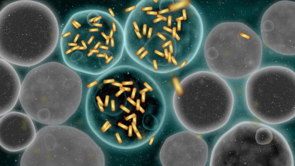
As Erica Locatelli, associate professor at Università di Bologna, explains nanorods are nanoparticles. They are small particles that have a nanometric size, made of gold and the shape of a cylinder. This allows the possibility to interact with light, and, in particular, with light that has no interference with the body absorption. This allows gold nanorods to be used in cancer detection and treatment, exerting some effects by laser irradiation.

However, gold nanorods, even if they are synthetised since many years, are not nowadays utilised in cancer treatment due to lack of reproducibility in synthesis and due to difficulties in scale up of their synthesis. That is something that must be addressed before producing nanorods for real cancer treatment.

Gold nanorods, however, are particularly challenging to produce at scale. For this reason, we have worked intensively on optimising critical parameters such as the preparation and storage of the seed solution, the addition of the growth solution, reaction conditions like peak temperature and time, and, most importantly, purification.

Purifying gold nanorods at this scale remains a major hurdle, as very few examples exist of nanostructured materials for diagnostic imaging being produced at such high volumes.

Addressing these technical challenges is essential for making gold nanorods a viable option in cancer diagnosis and therapy. By improving reproducibility and enabling reliable large-scale production, the PHIRE project is contributing to the advancement of this technology toward clinical use.

Enhancing precision in photothermal therapy

Photothermal therapy uses continuous laser irradiation to destroy cancerous tissue. A prediction map shows where the tumour is located, its size, and how long it needs to be irradiated to ensure complete eradication. This timing is calculated using complex mathematical models that simulate heat generation and transfer in tissue, as well as cell death due to rising temperatures.

However, these models are computationally heavy, often involving over a million equations, and require hours to solve, making real-time clinical application impractical.

To overcome this, the PHIRE team applies machine learning algorithms. By generating synthetic data from numerous scenarios (e.g. tumour size, position, laser power), they train AI models to rapidly estimate the ideal irradiation time for each patient. This enables clinicians to tailor treatment precisely and adjust the surrounding margin of tissue damage as needed, maximising tumour removal while protecting healthy tissue.